ABOUT US
GET A FREE QUOTE
-
Home

-
Products
-
Ultrasound Scanner
-
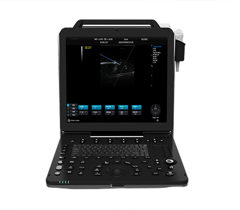
Trolley Color Doppler Ultrasound
- SM-90T 3D_4D_5D Trolley Digital Color Ultrasound Scanner
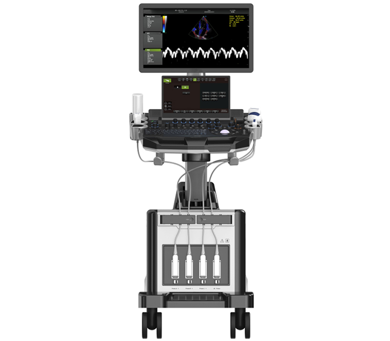
SM-90T 3D_4D_5D Trolley Digital Color Ultrasound Scanner
The high-end ultrasound applies outstanding image resolution, intelligence operation flow, ergonomic design and intimate man-machine interaction as an organic whole. High quality clinical performance, rich measurement software can quickly and conveniently solve the needs of cardiology, abdominal, peripheral vascular, superficial parts, skeletal muscle, gynecology, obstetrics, urology, newborn and other clinical diagnosis. Stable platform and user-friendly operation design improve the work efficiency of doctors to a greater extent.
-
Features
Continuous wave Doppler Imaging(CW)
Anatomic 3M Mode Imaging
Auto IMT(intima-media thickness) Measurement
3D/4D Imaging
Tissue Doppler Imaging(TDI)
Color M Mode Imaging
Trapezoidal Imaging & Panoramic Imaging
Free Hand Elastography
Rich Optional Transducers
- Trans-vaginal probe
- Convex probe
- Linear probe
- Micro-convex probe
- Phased array probe
- Trans-rectal probe
- 4D Volume probe
Technical specifications:
| High-end Color Ultrasonic Diagnostic Apparatus | |
| 1: | Summary of main specifications and system |
| 1.1 | Trolley type all digital color Doppler ultrasonic mainframe |
| 1.2 | Ultrasonic host operating system: Windows operating system |
|
1.3 |
Applications:
Abdomen, obstetrics, gynecology, heart, urinary system, small organs, superficial, blood vessels, pediatrics, newborns, musculoskeletal |
|
1.4 |
Probes:
Convex probe, Tran-vaginal probe, Linear probe, Micro-convex probe, Cardiac probe,4D Volume probe |
|
1.5 |
Applications and report:
Abdominal,OB,GYN,Cardiac,Urinary,Small Parts,Superficial, Vascular, Pediatrics, Advanced measurement software packages, report software packages, case management software packages, etc. |
| ☆1.6 | carotid artery intima measurement thickness(IMT) |
| ☆1.7 | Automatic spectral envelope measurement |
| 1.8 | Full digital transmission and reception of beam synthesizer |
| 1.9 | Color Doppler imaging(C) |
| 1.10 | Pulse Doppler Imaging(PW) |
| 1.12 | Continuous wave Doppler imaging(CW) |
| ☆1.13 | B/C/D Real-time three synchronous imaging |
| ☆1.14 | Power Doppler imaging(PDI) |
| ☆1.15 | Direct power Doppler imaging(DPDI) |
| 1.16 | M mode imaging |
| ☆1.17 | Anatomic M mode imaging |
| ☆1.18 | Color Doppler M mode imaging |
| ☆1.20 | Tissue Doppler imaging(TDI) |
| ☆1.21 | Strain rate imaging (SRI) |
| 1.22 | Tissue harmonic imaging(THI) |
| 1.23 | Fusion harmonic imaging(FHI) |
| 1.24 | Speckle Reduce imaging(SRI) |
| ☆1.26 | Deflection imaging |
| ☆1.27 | Trapezoidal imaging |
| 1.28 | Adaptive velocity optimization |
| ☆1.29 | Free hand 3D |
| 1.30 | Real time 3D imaging(3D/4D) |
| 1.31 | DICOM3.0 |
| 1.32 | Monitor:21.5 inch ,high definition ultrasonic display |
| 1.33 | 13.3 inch touch screen |
| 1.34 | Physical clipboard: save the image on the left side of the screen, which can be directly saved or deleted. |
| 1.35 | The system has the function of on-the-spot upgrade |
|
1.36 |
Presupposition: for different inspection of the viscera, preset the inspection conditions for the best image, reduce the adjustment of the operation, and the commonly used external adjustment and combination regulation. |
| 1.37 | Probe interface: 4 |
| 1.38 | Chinese and English System, Chinese and English input, optional |
| 1.39 | Depth:≥360mm; |
| 1.40 | Extended imaging |
| 2: | Probes |
| 2.1
Convex probe |
Fundamental Frequency:2.0MHz/2.3MHz/2.5MHz/3.0MHz/3.5MHz/4.0MHz/4.6MHz/5.0MHz/5.4MHz,
Harmonic Frequency:4.0MHz/4.6MHz/5.0MHz, |
| 2.2
Linear probe |
Fundamental Frequency:
4.0MHz/4.6MHz/5.0MHz/6.0MHz/7.0MHz/8.0MHz/9.2MHz/10.0MHz/12.0MHz/13.3MHz, Harmonic Frequency:8.0MHz/9.2MHz/10.0MHz, |
| 2.3
Trans-vagin al probe |
Fundamental Frequency:
3.0MHz/3.5MHz/4.0MHz/5.0MHz/5.4MHz/6.0MHz/7.0MHz/8.0MHz/10.0MHz, Harmonic Frequency: 6.0MHz/7.0MHz/8.0MHz, |
| 2.4 | Fundamental Frequency: |
| Micro-conv
ex probe |
3.0MHz/3.5MHz/4.0MHz/5.0MHz/5.4MHz/6.0MHz/7.0MHz/8.0MHz,
Harmonic Frequency: 6.0MHz/7.0MHz/8.0MHz, |
| 2.5
Cardiac probe |
Fundamental Frequency:
1.7MHz/1.9MHz/2. 1MHz/2.5MHz/3.0MHz/3.4MHz/3.8MHz/4.2MHz/5.0MHz, Harmonic Frequency: 3.4MHz/3.8MHz/4.2MHz, |
| 2.6
4D Volume probe |
Fundamental Frequency:
2.0MHz/2.5MHz/3.0MHz/3.3MHz/3.7MHz/4.0MHz/5.0MHz/6.0MHz, Harmonic Frequency: 4.0MHz/5.0MHz/6.0MHz, |
| 2.7:
Transrectal probe |
Fundamental frequency:
3.0MHz/3.5MHz/4.0MHz/5.0MHz/5.4MHz/6.0MHz/7.0MHz/8.0MHz/10.0MHz Harmonic frequency: 6.0MHz/7.0MHz/8.0MHz |
| 3: | 2D imaging mode |
| 3.1 | Gain:0-100 ,Step 2 adjustable |
| 3.2 | TGC: 8 segment adjustable |
| 3.3 | Maximum focus point:≥7, which can be moved throughout the whole process. |
| 3.4 | Speckle reduction:0-5 ,5 level |
| 3.5 | Space Synthesis:0-2 ,2 level(Liner probe: 3 level , cardiac probe:0) |
| 3.6 | Dynamic:30-180 ,35 level ,step 5 adjustable |
| 3.7 | Line density:low 、middle 、high ,3 level |
| 3.8 | Frame correlation:0-4,4 level |
| 3.9 | Noise reduction:0-5 ,5 level |
| 3.10 | Edge Enhancement:0-5 ,5 level |
| 3.11 | Sound power:2-10, 9 level |
| 3.12 | Grey scale:0-67, 67 level |
| 3.13 | False color:0-67 ,67 level |
| 3.14 | Image style: Soft-Comparison ,2 level |
| The screen has real-time display of voice power, probe frequency, dynamic range, pseudo color, gray scale and other 11 parameters can be adjusted | |
| 4: | Color Doppler imaging mode |
| 4.1 | Blood gain:0-100 ,Step 2 |
| 4.2 | Parameter display:Velocity、Variance |
| 4.3 | B-Restrain(B/W restrain):0-7, 7 level |
| 4.4 | Speed Through:0-8, 8 level |
| 4.5 | Sampling number:6-24, 7 level |
| 4.6 | Blood flow preferred:0-8, 8 level |
| 4.7 | Filtering: 1-6, 6 level |
| 4.8 | Sound power:2-6, 4 level |
| 4.9 | Noise reduction:0-4, 4 level |
| 4.10 | Smooth treatment:0-4 , 4 level |
| 4.11 | Frame correlation:0-6 , 6 level |
| 4.12 | Chromatography(Blood flow graph):0-37 , 37 level |
| 4.13 | Line density:Low-Middle-High , 3 level |
| 4.14 | Frequency:4 level adjustable |
|
4.15 |
Velocity:Minimum 0.4K ,Maximum 40.5K
Convex probe:0.4K-4.3K-38.5K Linear probe:0.4K-14.7K-39.0K Trans-vaginal probe:0.4K-7.8K-39.7K Volume probe:0.4K-4.2K-34.8K Micro-convex probe:0.4K-10.3K-40.5K cardiac probe:0.4K-7.8K-39.7K |
| PS:The frequency of the probe changes and the frequency value changes | |
| PS:Frame rate changes with speed | |
| 5: | Pulse wave Doppler(PW) |
| 1 | Gain:0-100 ,Step 2 |
|
5.2 |
Spectrum envelope function: real time automatic spectrum envelope, manual spectrum envelope, and other modes. The system automatically analyses and displays various data such as PSV, EDV, RI, PI, S/D, ACC, HR and so on. Can wake up or close |
| 5.3 | Sample volume:0.5mm~30mm |
| 5.4 | Blood angel:-75—75 degree ,Step 5 |
| 5.5 | False color:0-67 , 67 level |
| 5.6 | Dynamic range:20-40 , 4 level |
| 5.7 | Filter:0-9, 9 level |
| 5.8 | Smooth treatment: 1-4 , 4 level |
| 5.9 | Sound power:2-5 , 4 level |
| 5.10 | Volume:0-100, 10 level ,Step 10 |
| 5.11 | Audio filtering:0-4 , 4 level |
| 5.12 | Base line:-1.0~ 1.0, |
| 5.13 | Grey map:0-67 , 67 level |
| 5.14 | Scan velocity: 100-500, 6 level |
|
5.15 |
PRF:Minimum 0.5K ,Maximum 87.5K
Convex probe:0.5K-4.3K-63.3K Linear probe:0.5K-14.5K-78.4K Trans-vaginal probe:0.5K-8. 1K-78.4K Volume probe:0.5K-4.2K-53.8K Micro-convex probe:0.5K-10.3K-81. 1K cardiac probe:0.5K-4.3K-87.5K |
| 5.16 | Frequency:4 level |
| PS:The frequency of the probe changes and the PRF value changes | |
| PS:The frequency of the probe changes and the frequency value changes | |
| 6: | Continuous Wave Doppler(CW) |
| 6.1 | Support probe:Cardiac probe |
| 6.2 | Adjustment of B mode parameters is switchable |
| 6.3 | Gain:0-100 ,Step 2 |
| 6.4 | Sampling line position is adjustable |
| 6.5 | PRF:0.9K~36. 1K |
| 6.6 | Baseline:-1.0~ 1.0 |
| 6.7 | Blood angel:-75~75 degree |
| 6.8 | Grey map:0-67 |
| 6.9 | Scan velocity: 100-300 |
| 6.10 | False color:0-67 |
| 6.11 | Dynamic range:20-40 |
| 6.12 | Filtering:0-9 ,9 level |
| 6.13 | Smooth treatment: 1-4 |
| 6.14 | Frequency:2.0MHz/2.3MHz/2.5MHz/3.0MHz ,4 level adjustable |
| 6.15 | Sound power:2-5 |
| 6.16 | Volume:0-100 |
| 6.17 | Audio Filtering:0-4 |
| ☆7: | Anatomical M imaging |
| 7.1 | Support probe:Convex probe, Linear probe,Cardiac probe |
| 7.2 | Adjustment of B mode parameters is switchable |
| 7.3 | Gain:0-100 ,Step 2 |
| 7.4 | M Sampling line angel is adjustable |
| 7.5 | M Sampling line length is adjustable |
| 7.6 | Sampling line:3 ,Can be displayed or hidden separately |
| ☆8: | Blood flow M model(MC) |
| 8.1 | Adjustment of B mode parameters is switchable |
| 8.2 | Gain:0-100 ,Step2 |
| 8.3 | MC Sampling line angel is adjustable |
| 8.4 | MC Sampling line length is adjustable |
| 8.5 | Frequency:4 level |
| 8.6 | Sampling number:6-24 |
| 8.7 | Speed through:0-8, 8 level |
| 8.8 | Scan velocity: 150-500 |
| 8.9 | Frame correlation:0-6, 6 level |
| 8.10 | Filtering: 1-6 ,6 level |
| 8.11 | Blood flow preferred:0-8 ,8 level |
| 8.12 | Smooth treatment:0-4,4 level |
| 8.13 | Map:0-37, 37 level |
| ☆9: | Elastography |
| 9.1 | Adjustment of B mode parameters is switchable |
| 9.2 | Gain:0-100 ,Step 2 |
| 9.3 | B/E ,Double real-time display on the same screen |
| 9.4 | Probe displacement curve display:Up/Down |
| 9.5 | Pressure indicator bar display |
| 9.6 | Frequency: 8-9 level ,Adjustable;According to the probe display |
| 9.7 | Noise reduction:0-2, 2 level |
| 9.8 | Frame correlation:0-3, 3 level |
| 9.9 | Comparison:0-13, 13 level |
| 9.10 | False color:0-3 , 3 level |
| 9.11 | Don’t support cardiac probe |
| ☆10: | Tissue Doppler imaging(TDI) |
| 10.1 | Support probe: Cardiac probe |
| 10.2 | Adjustment of B mode parameters is switchable |
| 10.3 | Gain:0-100 ,step 2 |
| 10.4 | ROI area adjustable |
| 10.5 | Sampling number:6-24 |
| 10.6 | Velocity:0.4K-8.0K |
| 10.7 | Frame correlation:0-6 ,6 level |
| 10.8 | Tissue preferred:0-7, 7 level |
| 10.9 | Frequency:2.0MHz/2.3MHz/2.5MHz/3.0MHz |
| 10.10 | Support color reversal |
| ☆11: | Strain rate imaging |
| 11.1 | Support probe: Cardiac probe |
| 11.2 | Adjustment of B mode parameters is switchable |
| 11.3 | ROI area adjustable |
| 11.4 | Gain:0-100 ,Step 2 |
| 11.5 | Sampling number:6-24 ,6 level |
| 11.6 | Axial average: 1-4 , 4 level |
| 11.7 | Velocity:0.4K-8K |
| 11.8 | Frame correlation:0-6 , 6 level |
| 11.9 | Tissue optimization:0-7 ,7 level |
| ☆12: | Panoramic imaging |
| 12.1 | Support probe:Linear probe |
| 12.2 | Speckle Reduction:0-5, 5 level |
| ☆13: | Deflection imaging |
| 13.1 | Support probe:Linear probe |
| 13.2 | Adjustment of B mode parameters is switchable |
| 13.3 | Deflection angel: 8 level |
| 13.4 | Speckle reduction:0-5, 5 level |
| 13.5 | Dynamic rate:30-180 ,Step 5 |
| 13.6 | Line density:low-middle-high ,3 level |
| 13.7 | Frame Correlation:0-4 , 4 level |
| 13.8 | False color:0-67, 67 level |
| 13.9 | Image style: Soft-Comparison ,2 level |
| 13.10 | Noise reduction:0-5, 5 level |
| 13.11 | Edge Enhancement:0-5 ,5 level |
| 13.12 | Sound power:2-10 ,8 level |
| 13.13 | Grey map:0-67 ,67 level |
| ☆14: | Trapezoidal imaging |
| 14.1 | Probe support:linear probe |
| 14.2 | Adjustment of B mode parameters is switchable |
| 14.3 | Deflection angel: 8 level |
| 14.4 | Speckle reduction: 0-5, 5 level |
| 14.5 | Dynamic rate: 30-180 ,Step 5 |
| 14.6 | Line density: low-middle-high ,3 level |
| 14.7 | Frame Correlation: 0-4 , 4 level |
| 14.8 | False color: 0-67, 67 level |
| 14.9 | Image style: Soft-Comparison ,2 level |
| 14.10 | Noise reduction: 0-5, 5 level |
| 14.11 | Edge Enhancement: 0-5 ,5 level |
| 14.12 | Sound power: 2-10 ,8 level |
| 14.13 | Grey map: 0-67 , 67 level |
| 14.14 | Space Synthesis:0-2, 2 level |
| ☆15 | Freehand 3D imaging |
| 15.1 | Support probe:convex probe, linear probe |
| 15.2 | Display model: 4 pictures |
| 15.3 | Image Rotation X/Y/Z Axis |
| 15.4 | Multi-slice Visibility |
| 16 | Real-time 4D imaging |
| 16.1 | Support probe: 4D volume probe |
| 16.2 | Adjustment of B mode parameters is switchable |
| 16.3 | Gain:0-100 ,Step 2 |
| 16.4 | Display model:one image 、two images 、four images |
| 16.5 | Image Rotation:X/Y/Z Axis |
| 16.6 | Multi-slice Visibility |
| 16.7 | Light&Shade inversion |
| 16.8 | Smooth:0-4, 4 level |
| 16.9 | Threshold level:0-129, Step 3 |
| 16.10 | Transparency: 1-509 ,Step 10 |
| 16.11 | Render type:4 kinds ,Surface 、maximum 、minimum 、perspective |
| 17: | Extended Imaging |
| 17.1 | Gain:0-100 ,Step 2 |
| 17.2 | TGC: 8 segment adjustable |
| 17.3 | Maximum focus point:≥7, which can be moved throughout the whole process. |
| 17.4 | Speckle reduction:0-5 ,5 level |
| 17.5 | Space Compound:0-2 ,2 level(Linear probe: 3 level ,don’t support cardiac probe) |
| 17.6 | Dynamic range:30-180 ,35 level ,Step 5 |
| 17.7 | Line density:Low 、Middle 、High ,3 level |
| 17.8 | Frame correlation:0-4,4 level |
| 17.9 | Noise reduction:0-5 ,5 level |
| 17.10 | Edge enhancement:0-5 ,5 level |
| 17.11 | Sound power:2-10, 9 level |
| 17.12 | Grey map:0-67, 67 level |
| 17.13 | False color:0-67 ,67 level |
| 17.14 | Image style: Soft-Comparison ,2 level |
|
17.15 |
Extended level: Maximum 72 level
Convex probe:9 level Trans-vaginal probe:72 level Micro-convex probe:29 level Cardiac probe:40 level 4D Volume probe: 17 level |
| PS:The screen has real-time display of voice power, probe frequency, dynamic range, pseudo color, gray scale and other 11 parameters can be adjusted | |
| PS: When the probe scan range reaches the maximum, the space synthesized is 0. | |
| 18: | Measurement and analysis function: |
| 18.1 | General measurement: Distance, area, ellipse, cross line, angle, distance ratio, volume, Volume (ellipse), area ratio, diameter, joint angle |
|
18.2 |
Cardiac:Automatic spectrum envelope 、LV、Main Pulmonary artery diameter、RVEDd 、RVEDs、
LVM、LAV、HR、MVF、AO、AR、LVOT、TVF、Pulmonic valve、Pulmonary vein、RV、Doppler fetal heart sound、LVET、LVM、LVMI 、AV |
| 18.3 | Vascular: carotid intima (IMT), length stenosis ratio, area stenosis ratio, IMT (back wall), IMT (front wall) |
| 18.4 | OB:Fetal routine 、AFI 、TW 、GS 、CRl 、OFD 、HL 、ulna 、NT 、Fibula 、Nbonel、Radial 、Tibia |
| 18.5 | GYN:uterus、cervix、corpus uteri/cervix uterus、left ovarian vein、right ovarian vein、dominant follicle、 intima thickness |
| 18.6 | Urology: prostate、residual urine、left kidney、right kidney、left suprarenal vein、right suprarenal、left testis 、right testis 、left seminal vesicle 、right seminal vesicle |
| 18.7 | Abdomen:liver 、CHD、partal vein diameter、cholecyst 、CBD、pancreas 、spleen、Internal diameter of abdominal aorta 、kidney |
| 18.8 | Small parts:Thyroid |
| 18.9 | Software package:Measurement package 、Software package 、Medical records management software package |
| 19: | Graphic and text management system |
| 19.1 | Host build in 2 hard disk (SSD 128GB+HDD 1TB), Start fast and stable |
| 19.2 | Movie playback:≥1200 frames |
| 19.3 | Internal file information management system: can record patient number, name, check number, check date and so on, and can be searched and managed by numbering, checking number, name and so on. |
| 19.4 | Type of report is 16 |
| 19.5 | One key fast report graphic and text management |
| 20: | Interface |
| 20.1 | USB interface: 4 |
| 20.2 | HDMI interface: 1 |
| 20.3 | RJ-45 interface: 1 |
| 20.4 | Grounding wire interface: 1 |
| 20.5 | DVD RW: 1 |
| 21: | Configuration |
| 21.1 | Trolley type full digital color Doppler ultrasound diagnostic system main unit |
| 21.2 | Probe options: convex, linear , Trans-vaginal , phased array, micro-convex, 4D volume and trans-rectal |
| 21.3 | ≥13 quick adjusting knobs |
| 22: | Technology, after-sales service and other requirements |
| 22.1 | Two years warranty for both main unit and probes |
Related Products
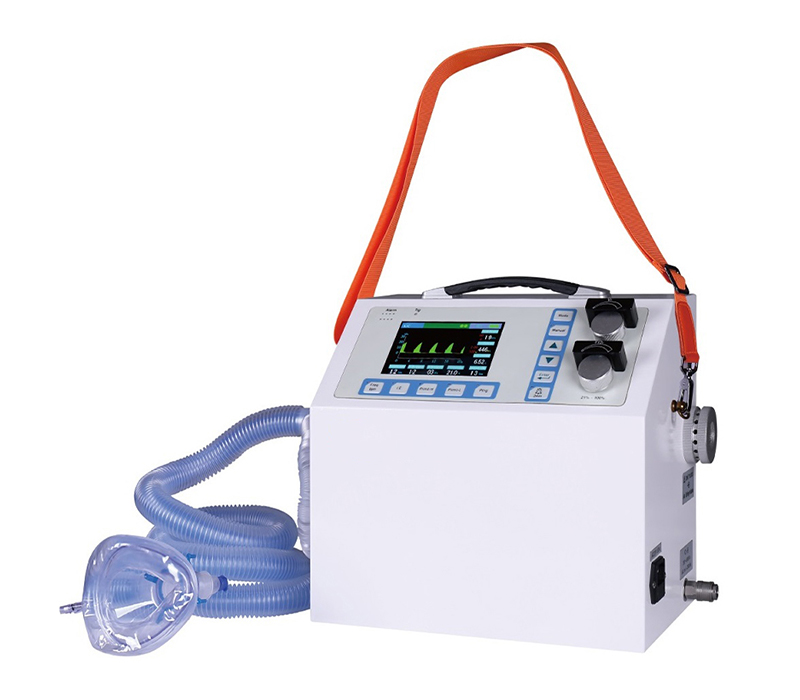
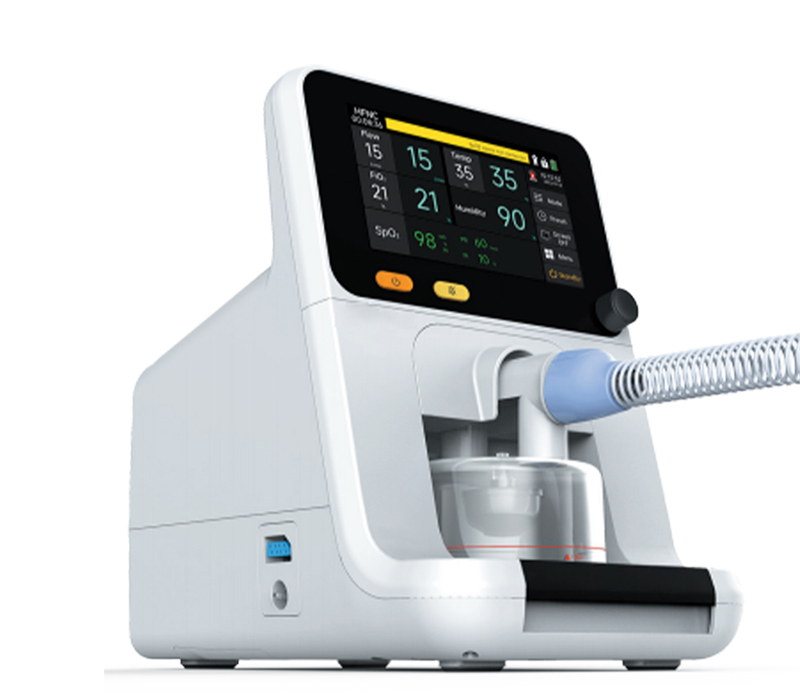
SMT-30F Medical Ventilator
High-flow nasal cannula
SMT-30F is a High Flow Nasal Cannula Oxygen Therapy Device capable of delivering up to 100% humidified and heated oxygen at a flow rate of up to 80L/min, it delivers a mixed oxygen with an easy controlled temperature and humidity. The oxygen supply system provides a great solution of airway management and oxygenation improvement.