ABOUT US
GET A FREE QUOTE
-
Home

-
Products
-
Ultrasound Scanner
-
Portable Color Doppler Ultrasound
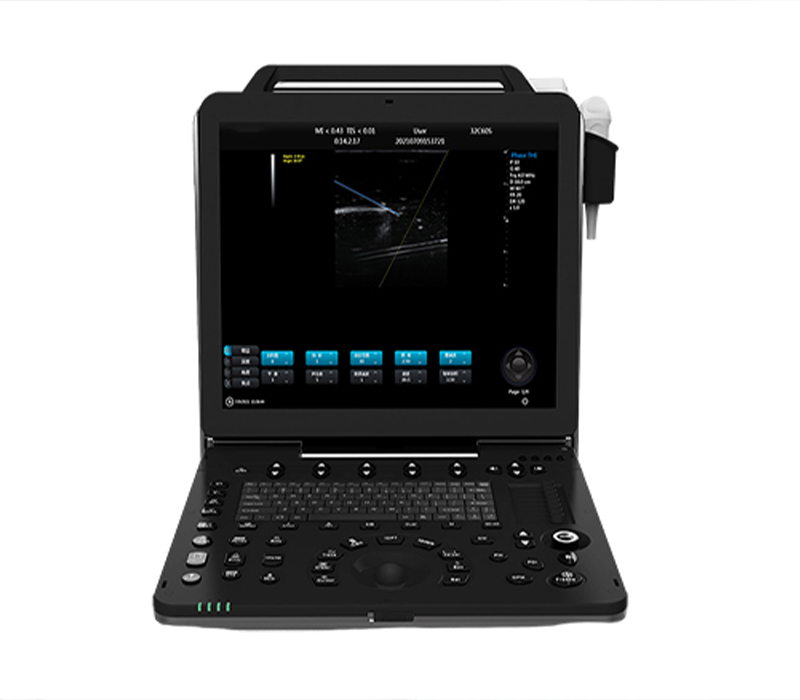
- SM-60P 3D_4D_5D Portable Full Digital Color Ultrasound Scanner

-
Features
4D real skin rendering function
Windows embedded ultrasonic running platform
Automatic spectrum measurement
The obstetric model can modify a variety of measurement combinations
Take ergonomic design into consideration
Hard disk dynamic and static image storage, real-time sharing
With automatic tracing, trapezoidal imaging, one key automatic optimization and other functions
DICOM function, can be directly connected with the hospital PACS system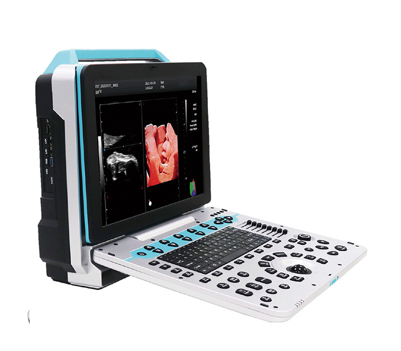
Technical parameters
- Product Name: Portable Color Doppler Ultrasound Diagnostic Instrument
1.1Structural type: portable
- Instructions for use and product requirements:
2.1 Meet the hospital’s examination and diagnosis needs in the areas of abdomen, obstetrics, gynecology, urinary system, small organs, superficial, vascular, pediatrics, neonatology, muscle, physical examination and other areas.
- Main specifications and system overview:
★ 3.1 Ultrasound host operating system: Windows 10 operating system
3.2 Spectral Pulse Doppler
3.3 Directional Energy Doppler
3.4 Real-time three-synchronization
3.5 Spatial composite imaging
3.6 Possessing tissue harmonic imaging technology
3.7 2B/4B Imaging Mode
3.8 System languages: Chinese, English, French, Russian and Spanish Portuguese, Indonesian, seven languages
3.9 Monitor: ≥15 inches
3.10 Integrated clipboard: Displays saved images on the left side of the screen and can be directly transferred or deleted
3.11 The system has the function of field upgrade
3.12 Preset conditions: Preset inspection conditions for optimized images for different inspections, reduce adjustments during operation, and commonly used external adjustments and combined adjustments
3.13 Probe interface ≥ 2
3.14 With trapezoidal imaging function
3.15 One-click intelligent optimization
- Probe:
1) Convex array probe
Frequency range: 2.0MHZ 2.5MHZ 3.0MHZ 3.5MHZ 4.0MHZ 5.5MHZ H4.0MHZ H5.0MHZ
Eight-level frequency conversion ( depth: 30 – 55mm)
2) Linear array probe
Frequency range: 6.0MHZ 7.5MHZ 8.5MHZ 10.0MHZ 12.0MHZ H9.0MHZ H10.0MHZ
Seven-segment frequency conversion ( depth: 20 -128mm)
3) Cavity probe
Frequency range: 4.5MHZ, 5.0MHZ, 6.0MHZ, 6.5MHZ, 7.0MHZ, 9.0MHZ, H7.0MHZ H8.0MHZ
eight-stage frequency conversion ( depth: 30 -156mm)
4) Phased Array Probes
Frequency range: 2.0MHZ 2.5MHZ 3.0MHZ 3.5MHZ 4.0MHZ 5.0MHZ H3.0MHZ H4.0MHZ
Eight-stage frequency conversion ( depth: 100 -244mm)
5) Volume probe
Frequency range: 2.0MHZ 3.0MHZ 4.0MHZ 4.5MHZ 5.5MHZ 6.0MHZ H5.0MHZ
Seven-segment frequency conversion ( depth: 30 -237mm)
6) Micro-convex probe
Frequency range: 4.0MHZ 6.0MHZ 7.0MHZ 8.0MHZ H8.0MHZ five-stage frequency conversion
( depth: 30 -111mm)
7) High-frequency phased array
Frequency range: 2.0MHZ, 3.0MHZ, 4.0MHZ, 5.0MHZ, 7.0MHZ, 5.0MHZ, 6.0MHZ, 7.0MHZ
Eight-stage frequency conversion ( depth: 100 -244mm)
8) Visual crowd probe
Frequency range: 4.5MHZ 5.0MHZ 6.0MHZ 6.5MHZ 7.0MHZ 9.0MHZ H8.0MHZ
Seven-segment frequency conversion ( depth: 30 -111mm)
9) Human rectal probe
Frequency range: 4.0MHZ 6.5MHZ 9.0MHZ H8.0MHZ Four-stage frequency conversion ( depth:
30 -110mm)
10) High-frequency linear array probe
Frequency range: 6.0MHZ 7.5MHZ 8.5MHZ 10.0MHZ 12.0MHZ H10.0MHZ
Six-stage frequency conversion ( depth: 20 -110mm)
- 2D imaging mode:
5.1 Gain: 0-100, step 1 Visible and adjustable
5.2 TGC: 8 adjustable levels
5.3 Dynamic range: 20-280dB 14 levels visually adjustable
5.4 False color: 0-11 levels, visually adjustable
5.5 Sound power: 5%-100%, 5% step, visually adjustable
★ 5.6 body position markers ≥ 80 types
5.7 Maximum number of focus points: 6 focus points, fully movable
- 8 Scan range: 50%-100%
- 9 screens with real-time display of sound power, probe frequency, dynamic range, pseudo color, grayscale and other 14 adjustable parameters
5.1 0: Scan line density: high, medium and low
- Color imaging mode:
- 1 Color flip: adjustable
6.2 B/C split screen synchronous display function: available
- 3 color baseline: 11 levels, visually adjustable
7 Spectral Doppler modes:
★ 7.1 Sampling volume angle correction: -80°~80°adjustable
7.2 Sampling volume: 0.5mm-20mm, visually adjustable
- 3 display layouts: ≥4 adjustable visual layouts
- 4 Speed scale: 32.8-328cm/s (different probes have different ranges)
★ 7.5 Spectrum envelope function: Real-time automatic spectrum envelope, manual
spectrum envelope and other modes are optional. The system automatically analyzes and
displays: PS, ED, PI, RI, S/D, HR and other data
★ 7.6 Automatic IMT measurement: Automatically detects intima-media thickness and
simultaneously analyzes the anterior and posterior walls of the carotid artery
8 3D/4D imaging modes:
8.1 Quick Angle: supports 0, 90, 180, 270° rotation of 3D window image;
8.2 Display layout: supports “double-frame”, “quadruple-frame” and “single-frame” image display;
★8.3 Reconstruction Mode: Five reconstruction modes: RealSkin, surface, Max, Min, and XRax;
8.4 X-axis, Y-axis, and Z-axis rotation support adjustable
★8.5 supports light source direction adjustment
9 Measurement and analysis functions:
9.1 Measurement items include distance, area, angle, time, slope, heart rate, speed,
acceleration, blood flow path, blood flow spectrum trace, resistance index/pulsatility
index and other professional measurements
9.2 Professional measurement packages are available according to the different organs
examined;
★ 9.3 The color and type of the measurement line can be adjusted at will (including
the activation color and the completion color)
★ 9.4 The display position and font size of the measurement results can be adjusted
as needed.
9.5 Professional software packages: abdomen, obstetrics, urology, etc.
- Image and text management system: Image saving format: BMP DCM JP E G
10.1 The host has a built-in ≥ 128G solid- state hard drive, which enables fast and
stable startup.
10.2 Movie playback: ≥600 frames
10.3 Built-in Archives information management system: can record number, name,
inspection number, inspection date, etc., and can search and manage by number,
inspection number, name, etc.
★ 10.4 Report types ≥16.
10.5 One-click quick report graphic management
10.6 has DICOM3.0 protocol and can be connected to PACS system
11 interfaces:
4 USB ports, 1 Audio, 1 HDMI, 2 RJ-45, 1 DP
12 configurations:
12.1 Portable color Doppler ultrasound diagnostic system host 1 unit
12.2 Probe: convex array probe (standard), linear array probe (optional), micro-convex
R15 probe (optional), cardiac probe (optional), cavity probe (optional), volume probe
(optional), etc.
12.3 Warranty for the whole machine 2 years
Related Products
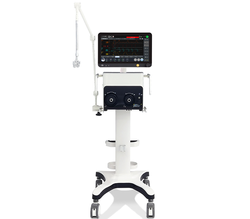
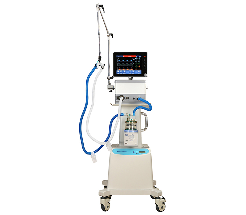
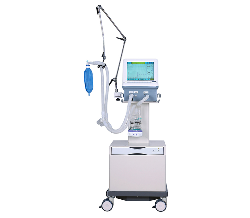
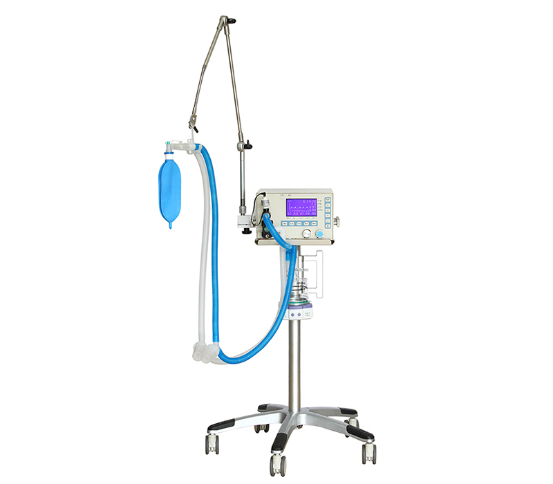
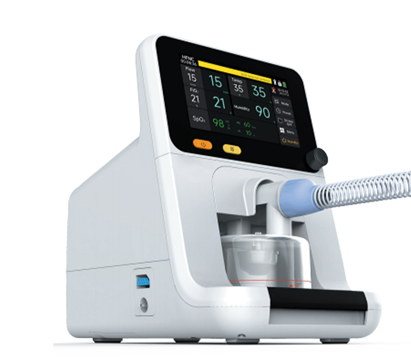
SMT-30F Medical Ventilator
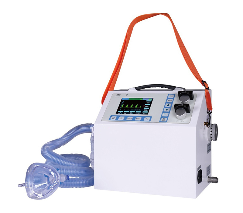
High-flow nasal cannula
SMT-30F is a High Flow Nasal Cannula Oxygen Therapy Device capable of delivering up to 100% humidified and heated oxygen at a flow rate of up to 80L/min, it delivers a mixed oxygen with an easy controlled temperature and humidity. The oxygen supply system provides a great solution of airway management and oxygenation improvement.